

Comparing laser eye surgery methods
It is possible to view laser eye surgery from two basic standpoints. Surgeries may be split into two groups – superficial or deep and standardised or individual. What are the differences and what is the optimal way of carrying out a surgery?
Superficial methods
These procedures come under many different names but in essence it is all about the removal or remodelling of the top layer of the cornea. The top layer of the cornea is called the epithelium and it is continuously being renewed. When removed, the nerve endings in the cornea are bared. This leads to considerable pain but for instance under contact lenses, the surface heals with a new layer of epithelium within 3–4 days. This regenerative characteristic of the surface of the cornea is made use of in all types of superficial surgeries. It is the epithelium that is manipulated with every time and the laser procedure is carried out beneath the epithelium. Which procedures are these?
- PRK (Photorefractive keratectomy)
- NO TOUCH
- LASEK
- EpiLASIK
It is an operating method whereby the epithelium is not removed mechanically but instead, in the first phase of the procedure it is removed using the excimer laser. The problem with this method is that in each patient, the epithelium is of a different thickness. It is impossible to measure this thickness upfront. The epithelium is therefore removed based only on an estimate and as such the final result often lacks precision. At our clinic, we do not perform this procedure.
This is a method whereby concentrated alcohol is used locally and is applied directly onto the epithelium of the cornea. Once peeled, a layer similar to the corneal lamella is created, however this one is usually formed by dead cells. Once the surgery is completed, this layer is replaced and functions to a certain extent as a contact lens does (which is, in most cases, applied to the eye anyhow). Paradoxically, this lamella, in some cases, slows down the healing process. At our clinic, we do not perform this procedure.
With this method, the epithelium of the cornea is folded using a special appliance. In such a case that an “untouched bridge” is retained, it is possible, once the surgery is completed, to replace and stretch out the epithelium into its original position. As compared to LASEK, the epithelium is more vital. Post-surgery covering using a contact lens is also common.
We do not perform the above described method at our clinic. Should the patient be unsuitable for the methods that we favour, namely the iDESIGN iLASIK or FemtoLASIK, we prefer to perform the PRK procedure because of its straightforwardness and relatively good predictability of the healing period. Another advantage of the PRK is the possibility to perform an individualised procedure (iDESIGN PRK). In such a case, however, we never operate both eyes simultaneously, instead we allow healing time in-between.
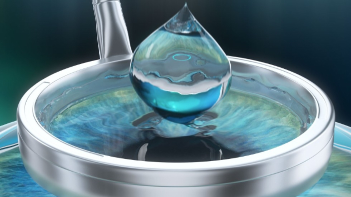
Lasek – application of alcohol on the epithelium of the cornea
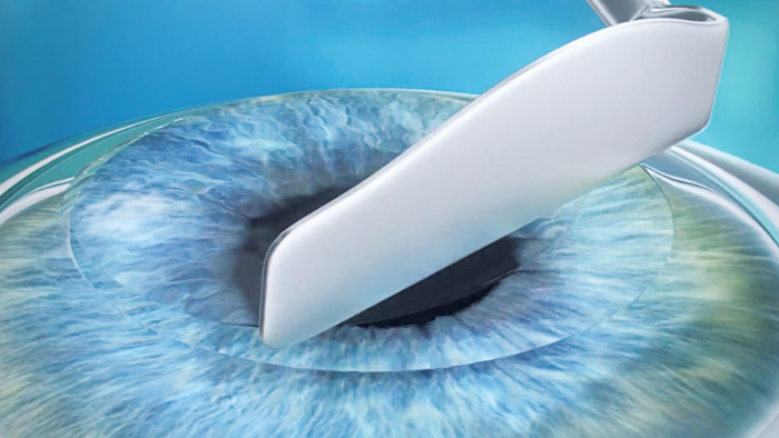
Lasek – uncovering the epithelium lamella
Deep surgeries
The common characteristic of the following methods is creating a corneal lamella, that copies the surface of the cornea and reaches into its deeper layer known as stroma. It is for this reason that these methods are often referred to as intra-stromal. Femtosecond laser is most commonly used nowadays to create the corneal lamella. In the past, microkeratome was used.
- LASIK (Laser in situ Keratomileusis)
- FemtoLASIK
- ReLEx Smile
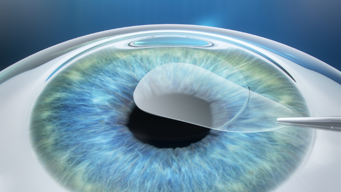
This method consists of the cutting away of a narrow lamella on the surface cornea (most commonly 100–140 microns thick) and subsequently creating a cornea bed using an excimer laser once the lamella has been lifted off. This lamella is returned back to its original position once the deeper layers of the cornea have been remodelled. It takes just a few minutes for the lamella to reattach and there is no need for sutures or covering the surface of the eye with a contact lens. The healing process of the eye after a LASIK procedure is very fast (just a few hours) and is painless. The post-surgery rehabilitation is more or less immediate and the eye is stabilised within a couple of days. This means only minimal limitations of the patient’s activities after the surgery.
The term LASIK is used to refer to a surgical procedure whereby a mechanical microkeratome is used to create a lamella. It is made up of two fundamental parts. The first part is attached to the surface of the eye so as to fixate it and prevent unwanted movement. The other part is then slid into the first part. It moves in a similar manner as a jack plane. A smooth cut is ensured by a fast moving (oscillating) razor blade. Safety and precision of the thickness and regularity of the lamella is ensured due to continuous improvement in technology. The success rate of this method is directly dependent on the dexterity and experience of the surgeon. Currently, this surgical procedure has been more or less eliminated because of the onset of the more modern and safer technology known as FemtoLASIK. This is why we no longer offer this procedure at our clinic.
It is a method using exclusively the energy of a femtosecond laser. It does not create a regular open lamella on the cornea but instead the femtoLaser creates a sort of 3D lens deep within the cornea. It is subsequently freed from the surrounding tissue and removed from the cornea using pincers through an incision of about 3 millimetres. The result is the thinning and flattening of the cornea at its centre. As such, this procedure is exclusively suitable for the correction of short-sightedness.
It is probably that this is the direction in which corneal laser surgery will be headed in the future. Firstly, however, the imperfections of this method must be eliminated. The most important problem with this method is its limitations in carrying out individualised procedures and removing for example irregular astigmatism. This is why, at the moment, the iDESIGN iLASIK method is currently by far the most superior. Another important and as yet unresolved issue is the difficulty of re-operating. Re-operating is sometimes the only option, especially in cases where the initial procedure was lacking in precision or when, after some time has passed, the dioptre error returns (we also operate young patients whose dioptre error may not be stabilised just yet).
These are but a few arguments why we do not, as yet, perform these surgeries at our clinic.
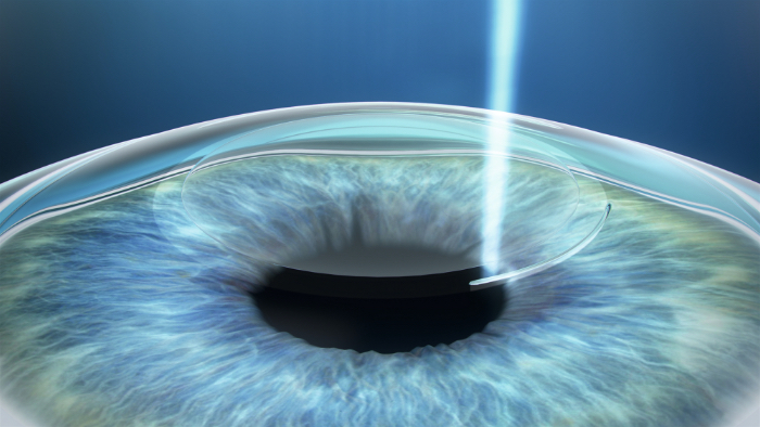
ReLEx Smile – creating a lenticule using Femtolaser
ReLEx Smile – extraction of corneal lenticule via a side incision
Fill out the appointment form and we will get back to you with suggestions of an appointment.
We further divide procedures into conventional and individualised
The conventional procedure refers to a surgery method whereby different patients with identical dioptre problem undergo an identical procedure. In this way, it is possible to easily eliminate common vision issues such as short-sightedness, far-sightedness or possibly a combination of the two or with symmetrical astigmatism. The remodelled surface is perfectly symmetrical similarly as glass used in regular glasses is. This technique ensures high quality post-op results and has been in use for many years.
What then is the individualised procedure and why is it recommended? In simple terms, this is a “tailor-made” procedure using state-of-the-art technology.
The basis for an individual procedure is the utilisation of super precise pre-operation analysis of the eye. Through such analysis, we measure the complexity of the impairment of each individual patient. Alongside problems with the axis (short-sightedness, far-sightedness and astigmatism) that may be removed using conventional procedures, this method has the potential to remove also higher-order abrasions and especially any irregularities that may be present. The next step is the preparation of a surgery plan with a proposed correction to the curvature of the cornea. Subsequently, the complete data is exported to the control unit of the laser.
This type of surgery is currently carried out using exclusively a lamella created through femto technology. The results of this method are perfectly precise. Once the lamella is lifted, a system enabling precise eye measurement is activated. For the entire duration of the operation, reference points on the iris are continuously being compared. These reference points were acquired during the patient’s pre-surgery examination (it is known as the iris map) and are scanned using a special camera during the laser procedure itself. This ensures absolutely precise incidence of the excimer laser rays on the surface of the cornea even when the patient continues to move his/her eye naturally. The quality of the laser ray itself is of utmost importance. Upon incidence, the ray must be capable of creating a perfect trail of different sizes. It is only in this manner that an absolutely perfect correction of the cornea and the removal of all optical imperfections may be achieved. All of the above factors, when combined together, ensure that the individual approach brings results exceeding 100% sharpness as is the case with for example the iDESIGN iLASIK procedure.