


Diagnostic instruments
iDESIGN
iDesign represents individualised and unique measuring of the optical properties of the eye. Modern refractive surgery deals not only with dioptre properties of the eye but also with parameters of the cornea, width of the pupil and wavefront analysis. Only flawless assessment of the above listed parameters enables us to receive the data that serve as basis for performing a precise laser eye procedure.
The iDESIGN instrument combines within itself the following diagnostic methods
- aberrometry (Wavefront analysis)
- automatic measuring of refraction
- pupillometry
- keratometry
- topography of the cornea
What then, makes the iDESIGN so unique? This type of analysis is currently considered to be the most precise of all. It enables us to collect all the necessary data during a single measuring and at the same time.
Using the data acquired, we create an algorithm to perform the laser eye surgery. Its subsequent realisation on the VISX Star device provides us with high quality pre-surgery results with vision sharpness exceeding 100%.
PENTACAM®
We use this device to examine the cornea and the anterior segment of the eye. The examination lasts only a few minutes, is contact-less and entirely painless.
It is the first diagnostic unit with automatically rotating Scheinpflug camera. During the rotational scanning, lasting approximately 2 seconds, up to 50 images of the anterior segment of the eye are captured. The examination is done automatically without any considerable strain on the patient.
Measuring parameters of the cornea
Pentacam® measures the cornea as a whole. It provides topographic data on the bulge and curvature of the anterior as well as posterior surface of the cornea. The thickness of the cornea (pachymetry) is measured and its entire surface is depicted graphically. The device is also capable of carrying out automatic recognition of the individual degrees of the keratoconus disease.
Other functions
For glaucoma screening, parameters such as the depth of the anterior chamber, the volume of the chamber (size) and the angles of the chamber are calculated and recorded. Illumination of the eye using the blue LED light makes clouding of the cornea and clouding of the lens (cataract) visible.
Once the Pentacam® examination is finalised, it draws up a report that summarises any abnormalities found during the screening.
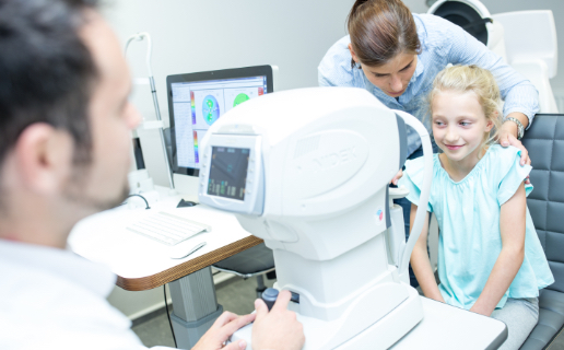
Examination using the auto-refraction keratometer
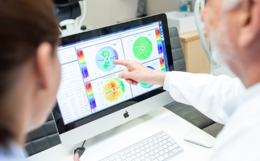
Examination using the Pentacam
Fill out the appointment form and we will get back to you with suggestions of an appointment.
Refractometer with keratometer and tonometer
Refractometer with keratometer is a device that decides the parameters of the eye including refraction. The patient places his/her chin on the base of the device and leans his/her head against the headrest. Thus, stability of the head is ensured as it is kept in a fixed position. Subsequently, the patient is asked to look into the device where he/she will see an image (hot air balloon) on which he/she is asked to focus. The eye relaxes and the device has time to make measurements.
The refractometer projects and image onto the retina and the image is reflected from the retina. It then evaluates the size and quality of the reflected image and it is based on this information that it calculates the refractive ability of the patient’s eyes. This is how we pre-determine the spherical dioptres and overall astigmatism.
As part of the so-called keratometry, we measure the dioptre refractivity of the cornea including detection of corneal astigmatism and determination of its axis.
This modern device thus determines the so-called objective refraction. This means that the patient’s parameters of the eyes are measured using an instrument unable to affect the measurement process. These, are, however only approximate parameters that assist the doctors or the optometrists to speed up and facilitate the examination of the subjective (patient’s own) refraction. This is an examination where using trial frames and lenses used in glasses, we determine the sphere and cylinder in such a way that the correction suits the patient and enables him/her to see as best as possible.
Contactless tonometry
It is the most commonly used examination method to measure intraocular pressure. It is performed using a contactless tonometer. The patient rests his/her head and chin and is asked to follow a colourful point inside the device. The device then blows three times into one eye and then three times into the other eye. The sharp gust of air flattens the cornea. It applies that the higher the intraocular pressure is, the more difficult it is to flatten the cornea. This is the basic principle of this measurement. Depending on the speed of the gust of air that is deflected, we calculate the pressure inside the eye based on a special algorithm.
Higher intraocular pressure may be falsely detected using this method when the patient has a thicker than average cornea or when the patient is clenching his eyes ever so slightly etc. This is why it is important to only use these results as indicative. This method is optimal for the screening for glaucoma.
Optical biometer NIDEK AL-Scan
Measuring axial length of the eye together with keratometry (measuring the refractivity of the cornea) serves as the basis for calculating of the optical capacity of the implanted intraocular lens according to various mathematical formulas. It is only thanks to precise measurements and the subsequent calculations that we are able to ensure the desired vision post cataract surgery and especially when carrying out refractive intraocular procedures.
Optical biometer NIDEK AL-Scan is not only fast but also extremely precise also in cases where there the pupil is severely cloudy. It enables 3D observation of the eye and automatic switching of the measurement process that goes unnoticed by the patient.
New mathematical formulas are continuously added and the so-called calculation A-constants of the different types of intraocular lens are thus optimised. The device also has special assistance available to enable correct adjustment of toric intraocular lens.