


Recurrent corneal erosion treatment
Recurrent Corneal Erosion is a condition where the cornea’s outermost layer of epithelial cells fail to attach to the underlying basement membrane (Bowman’s Layer). This is an unbearably painful condition because separation of these cells from the surface of the eye leads to exposure of the sensitive nerve endings in the cornea.
The symptoms include repetitive fits of acute pain, feeling a foreign body within the eye and photophobia (oversensitivity to light). The fits often occur immediately after waking up, during sleep (due to the spontaneous movement of the eyes) or when rubbing the eyes.
A recent damage to cornea (scratch or foreign body) is often discovered in the anamnesis. A cause may also be the dystrophy of the surface layers of the cornea. Erosion may appear also as a result of another eye disease.
The basic prevention is foregoing eye injury. We recommend using protective eye wear every time there may be a risk of eye injury. This is especially likely when working in the garden, specifically when handling leaves of plants, when trimming trees and shrubs etc… Similar injuries may come about when doing handcrafts where there is a risk of debris flying into the eye. Contact sports and playing with children are a chapter in its own right.
In case of erosion with primary disease of the cornea, the only preventive measures are, in fact, early diagnosis and subsequent adherence to a regime recommended by the doctor.
Preventive treatment
In an acute state, the best course of action is applying a protective contact lens and keeping it that way until the defect is completely healed over. Consequently, we recommend long-term systematic application of moistening eye drops during the daytime. Before going to bed, we recommend applying the same type of eye drops only with a higher viscosity – gel. The number of applications is entirely individual, most frequently this is 3–5x a day, with many patients having to apply the drops also during the night time.
With a deficiency of tears (dry eye syndrome), it is possible to use, as part of prevention, closure of the tear point using an implant. In case that we discover that the eyelids do not shut completely during sleep, we recommend using a night mask to cover the eyes.
Surgical treatment of recurrent erosion
- Alcohol abrasion
If a defect appears repeatedly on the surface of the eye (erosion) it is recommended to undergo the so-called epithelial abrasion.
This is a minor surgical procedure that is performed in an operating theatre under local topical anaesthesia. Having disinfected the area, ablation of the entire epithelium is performed using a surgical instrument (hockey stick). We then use the effect of concentrated ethyl alcohol to thoroughly cleanse the exposed surface including the bottom part of the actual defect. In this manner we prevent the formation of the so-called non-wetting corneal lesion that may form as a result of the effect of the greasy components of the tear film or after application of an eye ointment.
To complete the procedure, a contact lens is placed onto the treated cornea and we leave it that way for 2–4 weeks. The healing process is accompanied by a relatively intensive pain for at least the first 2–3 days. Once the contact lens is removed, the regular steps of preventing the formation of erosion apply – application of lubricants.
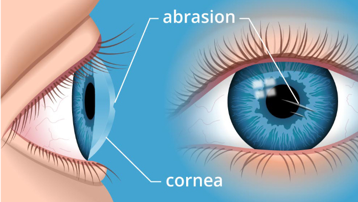
Recurrent corneal erosion
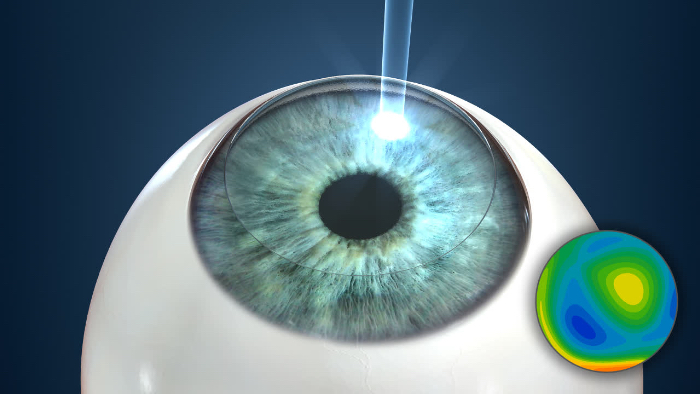
Photo-therapeutic keratectomy
Fill out the appointment form and we will get back to you with suggestions of an appointment.
- Anterior stromal micro-puncture
It is a procedure that is performed in the consulting room using a slit-lamp. This method is suitable in erosions placed within the periphery of the cornea. Under local anaesthesia, we perform surface perforations using a fine needle in the area of the separated epithelium. A more modern version of this method is the Nd: YAG laser micro-puncture, that uses laser pulses 0.4–0.5 mJ, applied to the area of the Bowman’s Membrane.
Based on great results, the idea to “grind-off” the surface of the cornea all over and thus secure a better and firmer attachment of the epithelium came about. To do so, instruments with a surface coarsened using diamond dust were used. Currently, usage of an excimer laser has replaced this technique (see below).
Under local anaesthesia, we perform surface perforations (punctures) using a fine needle. The punctures stimulate a reaction that lead to scarring between the epithelium and the surface layer of the cornea. These “bridges” help heal the defect in the epithelium.
- Excimer laser photo-therapeutic keratectomy (PTK)
This procedure is usually carried out under topical (surface) anaesthesia. Once the epithelium is removed, the patient actively co-operates by having to follow a fixed point. Only with eccentric or peripheral lesion the surgeon must manually turn the head or the eye of the patient or possibly use a joystick to place the laser beam in the desired spot.
PTK is carried out using an excimer laser (193 nm) and uses the principle of photoablation – the interruption the connection between molecules. A single pulse of the laser removes exactly 0.25 micrometres of corneal tissue. In this way, we have an option to plan ahead the exact desired change of the shape and ensure that the treated surface will be smooth and regular – this will improve the visual result. In case of treatment of the Recurrent Corneal Erosion the aim is to reach a dioptrically neutral result – meaning keeping the original dioptres. In such a case, the depth of the ablation on the entire surface, in the range of 6–9 millimetres, is only some 5–6 µm.
If the cornea is otherwise healthy, it is possible to combine the therapeutic effect PTK also with improved refraction. In such a case we are talking about carrying out PRK as PTK. In this way it is possible to eliminate myopia, possibly also myopia combined with astigmatism.
After the procedure we cover the surface of the cornea with a contact lens so that the pain is minimised. The pain following this type of surgery may be very intensive in the first 2–3 days post-surgery. We keep the contact lens in the eye for the duration of 2–4 weeks. The patient must, at the same time, apply antibiotic eyedrops. Once the contact lens is removed, the regular steps of preventing the formation of erosion apply – application of lubricants.