


Xantelasma
Xantelasma is a skin formation that is yellowish in colour and is usually occurs in the area of the eyelids. These formations are caused by the accumulation of fat and are painless. They may be a sign of an increased concentration of fats in the blood (hyperlipoproteinemia). This is why, if a person suffers from xantelasma – he/she ought to have their blood checked for the level of cholesterol and fat. This should not be underestimated as because a person with increased level of cholesterol and fat in the blood is also under a greater risk of a heart-attack. Xantelasma of different sizes, however, may also appear in completely healthy (usually older) individuals.
Treatment of xantelasma
Xantelasma is treated through extraction. The procedure is done under local anaesthesia. The incisions are made in the natural creases of the eyelids, in the crease of the upper eyelid and right beneath the eyelashes of the bottom eyelid. As a result, the incisions are practically invisible once healed. The incision is sutured after extraction and the stitches are removed after several days (7–10). The procedure is not difficult and as such the patient need not worry. This problem is recurring in many cases. Re-operation is an option
Of course, should it become evident that xantelasma occur due to hyperlipoproteinemia, it is necessary for the patient to go on a diet and other preventive measures may be recommended to lower the risk of heart-attack.

Xantelasma
Fibroma
Fibroma
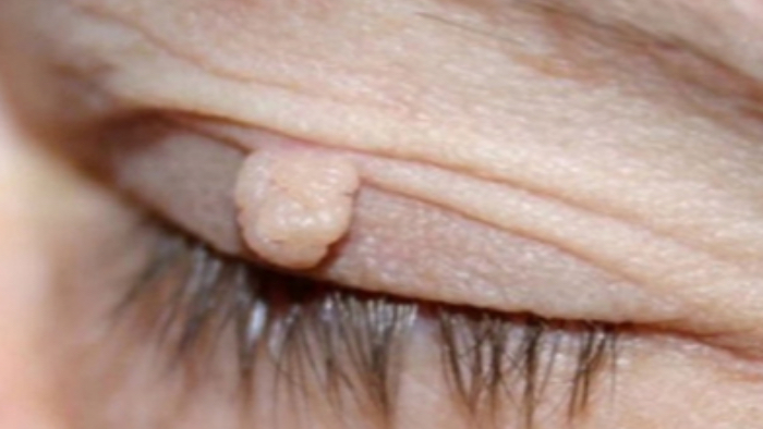
Fibroma (dermatofibroma) is a harmless round or oval growth that is red-brown in colour. It most commonly appears as an appendage on skin. It is painless and is similar in colour to surrounding skin or slightly darker. It occurs in various sizes ranging from miniscule to several centimetres large. It is usually round or oval-shaped and often is attached on a narrow stalk. The stalk may get twisted choking off the fibroma. As a result, blood supply cannot reach the fibroma causing painful reddish inflammation. The inflammation may spread out or the fibroma may fall off healing spontaneously. Fibroma is aesthetically damaging similarly as a wart is. It is easily removed and as such it is recommended to remove fibromas that get in the way, are painful, ugly or keep on enlarging.
Treatment of fibroma
We use two methods to treat fibroma – electrocauterization or excision.
- Electrocauterization
Electrocauterization means extracting tissue using thermos-destruction – meaning heat. The heat is generated using high frequency current. Using high frequency energy, it painlessly penetrates the tissue. The procedure is only very slightly painful, but it may be carried out under local anaesthesia.
The traces post-extraction are minimal and usually there is no scarring. Electrocauterization is very precise and removes the unwanted tissue without bleeding. Once extraction is carried out, a scab forms. It is advisable to not remove the scab but rather allow up to two weeks for it to heal spontaneously.
- Excision and suturing of the incision
The procedure is carried out under very strict sterile conditions under local anaesthesia. Larger incisions are covered using a bandage, smaller ones may be left uncovered. The healing period differs depending on the body part where the procedure took place. Usually, it takes 7–10 days, and this is when the stitches are also removed.
If necessary, it is possible to have the extracted tissue sent on for a histological analysis. This is one great advantage in comparison to laser removal.
Papilloma
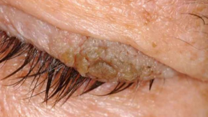
It appears on the skin of the eye lid as a cauliflower-shaped wart or a flat formation. There are usually several papillomata present at the same time. It is caused by the human papillomavirus and it can appear also in children. It is often recurring.
Treatment of papilloma
The treatment is surgical extraction of the formations using electrocauterization or excision. In recurring cases it is suitable to combine the excision with eventual cryotherapy (freezing) and administering of medication.
Papilloma
Verruca Vulgaris (wart)
Verruca Vulgaris (wart)
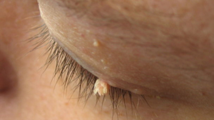
This is a problem caused by a virus infection on the skin. Warts are found all over the body but mostly on the hands.
Warts can be individual or multiple. Smaller daughter warts are usually found in the vicinity of the larger, so-called maternal wart. It is usually transmitted via direct contact, but indirect transfer is also possible (swimming pool, objects). The slightly raised bumps have a coarse surface formed by cracked keratin. The size may range from a few millimetres to several centimetres. As opposed to some other types of warts, malignant transformation usually does not occur with this type.
Verruca treatment
Therapy is complex. Even after surgical procedure, verruca often recurs. The individual lesions are repeatedly removed using electrocautery or surgical incision in local anaesthesia. In case of multiple verruca, what is of utmost importance is the extraction of the so-called maternal wart.
Atheroma
Atheroma is a skin cyst resulting from the blockage of a skin exocrine gland. It is usually accompanied by the over-production of sebum. The most common complication is its gradual enlargement (up to several centimetres) but more importantly, its purulence.
A surgical solution means freeing the contents of the cyst using a short incision and subsequent cleaning up using a surgical spoon then suturing the incision and if necessary, having a drain.
In cases with inflammation complications and when the atheroma is found in the face area a course of antibiotics is usually prescribed.