


Keratoconus
Keratoconus is a disease of the cornea that leads to its gradual thinning. This may cause blurred vision, double vision, worsened short-sightedness and astigmatism. Both eyes are usually affected, and sometimes scars and oedema may appear on the cornea besides the bulging. A fairly common accompanying symptom is allergy and rubbing of the eyes.
To date, the cause of this disease remains unknown. It is assumed that a combination of genetic and hormonal factors is to blame as well as the effect of the surrounding environment.
Keratoconus affects about 1 out of 2,000 people. The greatest incidence is in late childhood and early adulthood (15–20 years of age). It exists in all populations but is more frequent in people of Asian origin. About 7% of all those affected have a positive family anamnesis and thus heritability is probable.


Possible treatments of keratoconus
Non-surgical treatment options
Up to 90% of patients suffering from keratoconus will never require surgical treatment. We carry out treatment using the following types of contact lenses.
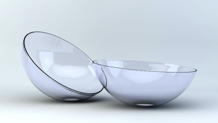
The advantage of soft contact lenses is the relatively good tolerance in wearing these. These lenses do, however, have insufficient effect in correcting higher degrees of symmetric as well as asymmetric astigmatism. As such exceptional vision is not to be expected.
- Rigid contact lenses
Rigid gas permeable contact lenses (RGP contact lenses) are the basic treatment of keratoconus. The lenses are relatively small, having a diameter from 8.0 to 9.6 mm. The principle is making use of the pressure force of the lens that limits the bulging of the cornea in an eye affected by keratoconus.
Rigid Contact lenses ensure relatively good vision and are affordable. Each lens is custom-made. Discomfort is a most common complaint of those wearing rigid lenses, given their small size and the fact that they move a few millimetres with every blink of the eye. The lens also transfers its entire weight onto the cornea which extremely sensitive, only adding to discomfort when worn. The movement of the lens when blinking also adds to unstable vision.
- Hybrid contact lenses
Hybrid contact lens is a combination of a small rigid lens (diameter of about 6 mm) and a large soft lens in one. Combining the positive characteristics of both types of lenses creates a single lens that is easy to put on and fits the eye well while ensuring good quality vision.
The disadvantages of hybrid contact lenses are a high cost and limited durability. The spot where the soft part joins the rigid part is problematic and separation of the two components happens frequently.
- Scleral contact lenses
Scleral lenses are designed so as to cover the entire surface of the cornea while at the same time so that they will not irritate the cornea through the pressure exerted on it. These lenses come into contact with the less sensitive part of the eye – the sclera (the white part of the eye). The size of the scleral contact lenses can be anywhere between 14.5 and 19.0 mm. The movement during blinking is minimal in these lenses (especially in comparison with rigid contact lenses).
When seeing this lens for the first time, most patients wonder how it at all is possible to insert it into the eye slit. Its size is what in most cases in limits its usage. Those who are able to insert it on the other hand, praise its comfort and especially the quality of vision.

Surgical treatment options
- Corneal Collagen Cross Linking
Collagen Cross Linking (CXL) method includes saturating the tissue of the cornea with solution of riboflavin. Once the riboflavin penetrates the cornea, the tissue is exposed to UV rays. The riboflavin (activated by the UV radiation) considerably increases the number of connections between the collagen fibres (cross linking). Increased solidity and stabilisation of the cornea is thus achieved, and this makes it possible to prevent its further steepening.
CXL is most suitable for younger patients but it may positively help patients of all age groups where the keratoconus remains unstable. Besides the increased solidity of the cornea, in some case the cornea also becomes flatter post-surgery.
CXL is considered to be a safe method. The most common complication is pain lasting for up to several days after the procedure. Some 25% of patients mention considerable pain, 45% mention no pain at all. Most patients register post-procedure discomfort in the form of dry eyes. Progressive bulging of the cornea has been halted in some 98% of all cases where keratoconus was treated using the CXL. A haze lasting more than 6 months was mentioned in less than 2% of all patients.
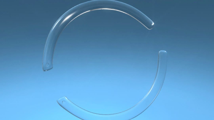
A surgical procedure with implantation of corneal segments is more difficult. In its first phase a tunnel is made of a 2 thirds depth into the cornea outside the pupil. The size and shape correspond to that of the implant to be used. Nowadays, femtosecond laser is used exclusively to create the tunnel. As soon as this tunnel is created, a plastic segment corresponding to a circular cut-out of about is 180° to 360° is inserted. Up to two segments may be implanted at one time.
The principle of implanted segments lies in their centrifugal pull onto the surrounding tissue. This leads to flattening of the central part of the cornea with the keratoconus. Patients may subsequently find wearing of rigid contact lenses problematic. Correction using soft contact lenses is usually without problems.

- Corneal transplantation
Up to 5% of all patients with keratoconus will undergo corneal transplantation in the future. This means that the cornea of the patient in question will be replaced by a cornea from a donor. An exchange of 100% of the tissue is known as penetrative keratoplasty (PK, PKP). A modified version of the transplantation of the cornea is a method known as DALK (Deep Anterior Lamellar Keratoplasty). With DALK only the outside parts of the cornea are replaced while the inner layers of the cornea (Descemet membrane and endothelium) remain intact.
If the reduced sharpness of vison is primarily a result of scar tissue of all the layers of the cornea of (or in the area of the Descemet membrane) then penetrative keratoplasty is performed. Usually though the scar tissue in patients is found mostly in the outside layers. The DALK method is more beneficial in such a case.
Patients suitable for transplantation of the cornea have usually undergone several of the above-mentioned methods in the past, but unfortunately with less than satisfactory results. Because transplantation replaces the affected (bulging cornea) using new tissue, the result is normally a better curvature of the cornea. A new, clearer and more symmetrical cornea ensures improved vision. Having undergone this type of surgery, it is possible to keep on improving the quality of the achieved vision using glasses or contact lenses.
The most common complication with transplantation of the cornea are:
- infection
- rejection of the donor cornea (some 3 to 30% depending on the method)
- asymmetrical astigmatism and a residual refractive error
- complications with usage of contact (especially soft ones)