


Corneal Ecstasy is a disorder where the cornea becomes thinner. This leads to the bulging of the cornea, the emergence of an asymmetrical astigmatism or, in rare cases, its perforation. Keratoconus, PMD (Pellucid Marginal Degeneration) or Keratoglobus are among these diseases, where the cause still remains unknown. Ecstasy of the cornea may appear also after a previous laser eye surgery.
Ecstasy diseases of the cornea may affect vision, but it may be renewed using optical aids such as glasses and soft as well as firm (gas permeable) contact lenses. Serious cases, however, may require a correction or renewal of the tectonic integrity of the cornea surgically. Earlier, PKP (penetrating keratoplasty) was a form of treatment. Even though PKP may be successful in patients with this diagnosis, the healing process after PKP may include a high post-surgery astigmatism, side effects of the used local steroids, rejection of donor cornea etc.
A new possibility in the surgical treatment of ecstasy diseases of the cornea is the implantation of Intracorneal Ring Segments – (ICRS).
These are usually manufactured from PMMA (polymethylmethacrylate) and are implanted outside the central optic zone – 4 millimetres into the tunnel created inside the deeper spheres of the corneal stroma, to be exact. Intracorneal Rings were initially meant for the treatment of short-sightedness, later, this method was also recommended for the treatment of keratoconus. It was discovered that using this method prevented further bulging of the cornea and could lower astigmatism associated with keratoconus.
There are different types of ICRS on the market. These differ in their geometrical profile and the diameter of their curvature. For example:
- INTACS
- Ferrara Rings
- Myoring
Surgical procedure
The first step is the plan of the procedure. A thorough analysis of the patient’s data acquired in the initial examination at the corneal advisory centre is carried out. An individual procedure according to the company’s regulations and expertise of the workplace is put forward. An optimal type of ICRS is determined (shape, curvature and thickness of the segment, how many degrees of the circular segment, one or two segments) so that optimal post-surgery results may be achieved.
The surgical procedure itself is carried out under topical (surface) anaesthesia. The centre of the pupil or the geometric centre of the cornea is marked on the cornea. The tunnels and the entry incisions are made exclusively using a femtosecond laser. Using this technology enables us to make tunnels of precise shape and in the exact required depth. The femtosecond laser software enables us to make custom-made tunnels and incisions.
The selected segments are then carefully inserted into the prepared tunnels using special. At the end of the surgery a contact lens is placed onto the eye to cover it until the next day.
This procedure is often simultaneously combined with Collagen Cross Linking (CXL) at some clinics. Other clinics do not perform CXL at all and other clinics still recommend performing CXL only after 4 to 6 months.

A couple of segments in the corneal tunnel with flattened central part of the cornea
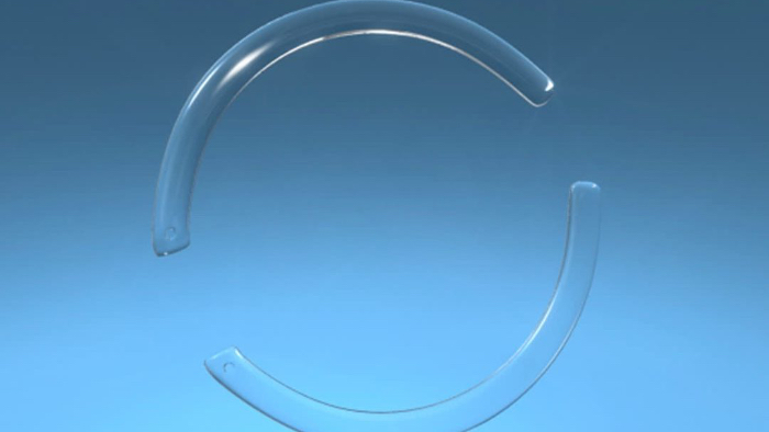
Corneal segments with a cut out sector of 170 degrees
Fill out the appointment form and we will get back to you with suggestions of an appointment.
Complications
Complications are rare because the implantation of the ICRS is reversible, no corneal tissue is extracted during the procedure and the central optical zone remains unaffected. Most potential complications were removed with the onset of the use of the femtosecond laser. Serious risks such as surface dissection of the canal, decentralisation of the tunnels or perforation of the frontal chamber were thus eliminated.
Later post-surgery complications were also reduced – for example epithelial growths and healing disorders in the place of the entry incision, extrusion of the segments, neovascularisation of the cornea, migration of the segment (sudden unexpected movement), infection of the cornea (keratitis), canal deposition (foreign body fragments), opacity of the cornea, dissolving of the cornea, chronic pain etc.
Results
In all studies, a significant central flattening of the cornea has been recorded. It is possible to expect stabilisation of the result in about 1 to 6 months post-surgery. The positive effect of the ICRS further supports the fact the most studies proved an improvement in the best visual acuity corrected using glasses (BSCVA) in 50% of all cases. It was also established that ICRS is helpful in increasing tolerance towards contact lenses on the eye used for the correction of the residual refractive error. Implantation of ICRS is thus a safe and successful alternative in treatment of keratoconus, PMD and post-laser ecstasy.
Who is eligible for this method?
ICRS may be used in the treatment of short-sightedness (from −1.00 to −3.00 dioptres) in patients with keratoconus and with ecstasy after refractive laser procedures. Indication for the ICRS implantation is transparency of the central part of the cornea and intolerance to wearing contact lenses. The cornea should be at least 400 μ thick in the place where the tunnel is intended.
Contraindication
ICRS should not be used in patients with keratoconus who may achieve functional daily vision using contact lenses and those that have no problems in wearing regular contact lenses. Furthermore, also in those who have non-transparent central part of the cornea and where the thickness of the cornea in the place of the intended tunnel is less than 400 μ.